|

|

|

|
Rice Dwarf Virus outer shell, inner shell, segmented compoment of outer shell,
and segmented compoment of inner shell.
The figures appear in project
3D Cryo-Electron Microscopy.
|

|

|

|
(a)
|
(b)
|
(c)
|

|

|

|
(d)
|
(e)
|
(f)
|

|

|

|
(g)
|
(h)
|
(i)
|

|

|
(j)
|
(k)
|
3D cryo-EM image of Rice Dwarf Virus (RDV) processed via our meshing pipeline.
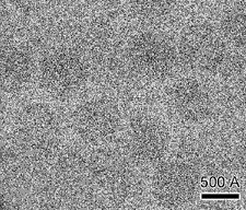
(a): two dimensional projection of an acquired cryo-EM image of the virus particles.
(b): reconstructed 3D image of intensities -bisected to show interior and volume rendered.
(c): the RDV capsid with inner core packing of proteins and nucleic acids,
volume rendered with appropriate opacity transfer function.
(d): RDV shown in (c) after contrast enhancement step.
(e): segmentation before filtering into an outer capsid layer (blue),
inner capsid layer (purple) and the inner core (green). Note noise around the outer capsid.
(f): segmentation applied after anisotropic filtering of the 3D image.
(g): outer capsid layer is further automatically segmented into locally asymmetric subunits.
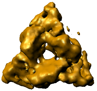
A P8 trimeric subunit is identified.
(h): surface rendering of the segmented P8 trimeric subunit from the outer capsid.
(i): the P8 trimeric subunits is further segmented into three P8 monomeric proteins (colored differently).
Triangular surface (j) and tetrahedral volumetric mesh (k) of each P8 monomeric protein subunit are shown.
|
|


