Although atomic details are not detectable in reconstructed 3D cryo-EM maps, given their low feature
resolution, it is sometimes feasible to locate secondary structures (alpha helices
and beta sheets) [1]. An approach for detecting alpha helices in 3D maps is where the alpha helix
is modelled with a cylinder (length and thickness) and the cylinder is correlated with
the segmented protein map. Since the best solution is achieved by exhaustively searching in translation
space (3D) and orientation space (2D), this method is computationally expensive. A
related exhaustive search approach, designed for beta sheet detection uses a disk (planar) model for
beta sheets. To detect secondary structures efficiently one must avoiding an
exhaustive search in both translation and orientation space. One possible approach is to consider
scoring candidate helices/sheets only at the Morse critical points of the 3D Map,
thereby reducing the search in translation space to a significantly smaller number of locations.
In addition, the search in orientation space at each critical point can be further
reduced by utilizing the local structure tensor. Using a criterion based on the eigenvalues of the
local structure tensor one is able to distinguish between alpha helices (line features)
and beta sheets (plane features). A critical point classified as an alpha helix, is extended on both
sides along the direction of the line structure determined by the local structure
tensor, yielding a segment of the median axis of the 3D map. Similarly, for a critical point
corresponding to a beta sheet feature, the plane feature is extended yielding a piece of
median surface of the density map. Since a true alpha helix or beta sheet may consist of more
than one critical point, it is necessary to merge a number of median segments and median
surfaces, from which the final alpha helixes and/or beta sheets are constructed.
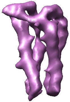
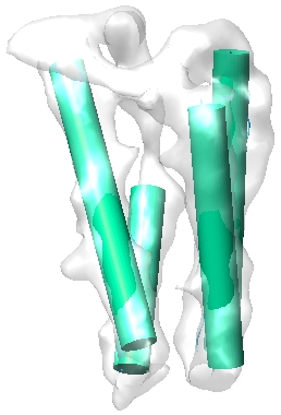
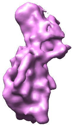
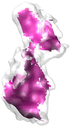
Shown below are examples of the secondary structure detection from blurred maps of crystal structures at 8 angstroms.
 |
 |
 |
|
PDBID = 1BBH (left: Contoured Blurred 3D Map, middle: Detected helices (green) from 3D Map; right: Helices (green) from PDB structure) |
 |
 |
 |
|
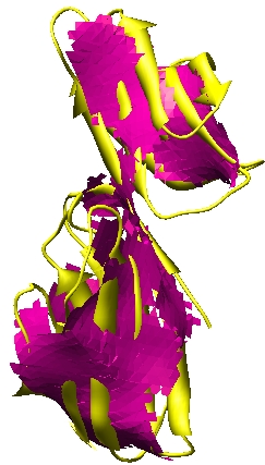
PDBID = 1CID (left: Contoured Blurred 3D Map, middle: Detected Sheets (pink) from 3D Map; right: Sheets (pink) from PDB structure) |
 |
 |
 |
|
PDBID = 1IRK (left: Contoured Blurred 3D Map, middle: Detected Helices (green) and Sheets (pink) from 3D Map; right: Helices (green) and Sheets (pink) from PDB structure) |
Publications
- Chandrajit Bajaj and Zeyun Yu, "Geometric Processing of Reconstructed 3D Maps of Molecular Complexes",
in Handbook of Computational Molecular Biology, Edited
by S. Aluru, Chapman & Hall/CRC Press, Computer and Information Science Series, October 2005.
|


